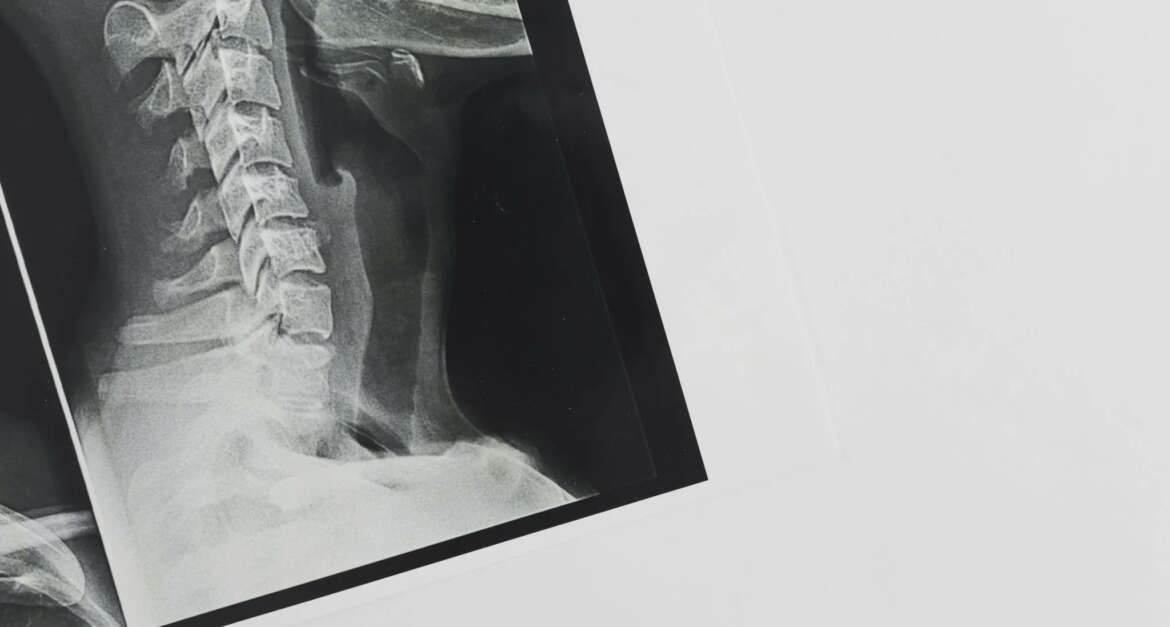
Neck fractures are serious injuries that require a prompt and accurate diagnosis for appropriate treatment. The neck, or cervical spine, plays a crucial role in supporting the head and protecting the spinal cord. In this blog post, we will provide a comprehensive guide to the diagnosis and imaging techniques used in identifying neck fractures. We will also highlight the expertise of Dr. Vivek Bonde, a renowned spine and brain surgeon, who specializes in the diagnosis and treatment of neck fractures. For more information about Dr Bonde and his practice, please visit his website at https://www.spinenbrain.com/.
Understanding Neck Fractures-
Neck fractures can occur as a result of various traumatic incidents, such as car accidents, falls or sports injuries. They can range from mild compression fractures to more severe fractures that involve displacement or instability of the cervical spine. Prompt diagnosis is crucial to prevent further damage to the spinal cord and ensure appropriate treatment.
Clinical Assessment-
When a patient presents with a suspected neck fracture, a thorough clinical assessment is the first step in the diagnostic process. A skilled spine specialist like Dr Vivek Bonde will conduct a comprehensive evaluation, which includes assessing the patient’s medical history, conducting a physical examination and evaluating neurological function. This evaluation helps identify signs and symptoms that may indicate a neck fracture and guide further diagnostic investigations.
Imaging Techniques for Neck Fractures-
a. X-Rays- X-rays are often the first imaging technique used to evaluate neck fractures. They can provide valuable information about the alignment, integrity and potential fractures of the cervical spine. Dr Bonde’s website provides detailed information about the importance of X-rays in diagnosing neck fractures and their role in guiding treatment decisions.
b. Computed Tomography (CT) Scan- CT scans are highly effective in assessing the bony structures of the neck and providing detailed images of potential fractures. They can reveal fractures that may not be visible on X-rays and help determine the extent and severity of the injury. Dr Bonde utilizes advanced CT imaging techniques to obtain precise three-dimensional images of the cervical spine, allowing for accurate diagnosis and treatment planning.
c. Magnetic Resonance Imaging (MRI)- MRI is particularly useful in evaluating the soft tissues surrounding the cervical spine, including the spinal cord, nerves, and ligaments. It can help identify associated injuries, such as spinal cord compression or nerve damage. Dr Bonde’s website highlights the role of MRI in providing a comprehensive assessment of neck fractures and its significance in guiding surgical interventions, if necessary.
Dr Vivek Bonde’s Expertise in Neck Fracture Diagnosis
Dr Vivek Bonde, a highly skilled spine and brain surgeon, specializes in the diagnosis and treatment of neck fractures. His website, https://www.spinenbrain.com/, provides valuable insights into his experience, qualifications, and commitment to patient-centred care.
Dr Bonde’s extensive expertise allows him to accurately diagnose and classify neck fractures, guiding the appropriate treatment approach for each patient. His multidisciplinary approach involves close collaboration with radiologists, physical therapists and other healthcare professionals to provide comprehensive care.
Conclusion
Diagnosing neck fractures requires a systematic approach and the use of advanced imaging techniques. Dr Vivek Bonde’s expertise and dedication to patient care make him an invaluable resource for individuals with neck fractures. By visiting his website, https://www.spinenbrain.com/, patients can access detailed information about neck fracture diagnosis and the range of treatment options available. With Dr Bonde’s commitment to staying at the forefront of medical advancements and his patient-centred approach, individuals with neck fractures can trust that they are receiving the highest standard of care.

